Recent neurological research has uncovered a dynamic and evolving response within the brain following peripheral nerve trauma, suggesting that the neurological impact of chronic pain is far more systemic and progressive than previously understood. A study conducted by Cazuza et al., published in the January 2026 issue of the Journal of Pain, utilized advanced automated mapping to track the behavior of microglial cells in rats following the induction of sciatic nerve injury. The findings demonstrate that neuroinflammation is not a static reaction but a shifting process that migrates across various functional regions of the brain as an injury transitions from an acute to a chronic state. This discovery provides a high-resolution view of how the central nervous system adapts to prolonged distress, potentially explaining the complex comorbidities often associated with chronic pain, such as sleep disturbances, emotional volatility, and cognitive decline.
The Role of Microglia in Neurobiological Maintenance
To understand the significance of the study, it is essential to define the role of microglia within the central nervous system. Microglia are the primary immune cells of the brain, distinct from other glial cells such as astrocytes and oligodendrocytes. While neurons are responsible for transmitting electrical signals, microglia serve as the resident macrophages of the brain and spinal cord. They comprise approximately 10% of the total cell population in the brain and are responsible for a wide array of homeostatic functions, including the pruning of redundant synapses during development, the clearance of cellular debris, and the primary response to pathogens or trauma.
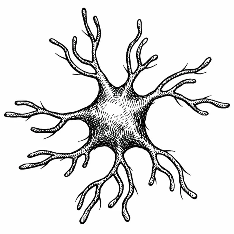
Under normal physiological conditions, microglia exist in a "surveying" state, characterized by a small cell body and long, highly mobile processes that constantly probe the surrounding environment. When they detect signals of injury or inflammation, they undergo a rapid morphological transformation, shortening their processes and enlarging their cell bodies—a state often referred to as "activation." While this response is necessary for immediate repair and protection, prolonged microglial activation has been linked to various neurodegenerative conditions and the maintenance of chronic pain states through the release of pro-inflammatory cytokines.
Study Methodology and Chronology of Nerve Injury
The research team led by R.A. Cazuza sought to map these microglial changes with unprecedented scale and precision. The study utilized a rat model of sciatica, induced through a controlled constriction of the sciatic nerve, which mimics the mechanical compression often seen in human radiculopathy. To ensure a comprehensive overview of the brain’s response, the researchers analyzed 52 distinct brain regions using automated morphometric software. This technology allowed for the objective measurement of thousands of cells, providing a statistical density of data that was previously unattainable through manual observation.
The study focused on two critical time points: 7 days and 28 days post-injury. These intervals represent the transition from the sub-acute phase of nerve injury to the establishment of chronic neuropathic pain. By comparing the morphological state of microglia at these two stages, the researchers were able to plot a timeline of neurological adaptation.
The Initial Phase: Day 7
One week after the nerve injury, the most significant microglial changes were concentrated in the motor cortex. This region of the brain is primarily responsible for the planning and execution of voluntary movements. The activation of microglia in this area suggests that the brain’s immediate response to peripheral nerve damage is heavily focused on motor adaptation and "guarding" behaviors. As the animal adjusts its gait and posture to compensate for the sciatic pain, the motor cortex undergoes a period of high plasticity and inflammatory signaling.
The Chronic Phase: Day 28
By the 28th day, the landscape of microglial activation had shifted significantly. While the initial motor cortex activity began to stabilize, new patterns of activation emerged in the hypothalamus and the brain’s reward circuitry, including areas involved in motivation and stress regulation. This temporal shift is highly significant, as it mirrors the clinical progression seen in human patients, where long-term pain ceases to be a purely sensory experience and begins to manifest as a psychological and systemic burden.
Supporting Data and Functional Mapping
The data revealed that microglial activation was not confined to the somatosensory pathways—the areas of the brain that traditionally process "where" and "how much" it hurts. Instead, the "whole-brain" involvement suggests that chronic pain recruits networks responsible for emotion and cognition.

The involvement of the hypothalamus at the 28-day mark is particularly noteworthy. The hypothalamus acts as the command center for the autonomic nervous system and the endocrine system, regulating sleep cycles, appetite, and the stress response (the HPA axis). Microglial activation in this region provides a biological basis for why chronic pain patients frequently suffer from insomnia and heightened stress sensitivity.
Furthermore, the activation observed in the reward circuitry—specifically regions involved in dopamine signaling—points toward the neurological roots of pain-induced depression and anhedonia (the loss of interest in pleasurable activities). When the brain’s reward centers are in a state of neuroinflammation, the ability to process positive reinforcement is diminished, leading to the "emotional exhaustion" often reported by those with long-term sciatica or similar nerve injuries.
Analysis of Implications for Human Medicine
While the study was conducted on rodent models, the implications for human clinical practice are substantial. The research reinforces the "biopsychosocial" model of pain, which posits that chronic pain is a complex interaction of biological, psychological, and social factors. By demonstrating that the brain literally changes its inflammatory profile over time, the study provides a tangible target for future therapeutic interventions.
Current treatments for sciatica and neuropathic pain often focus on the site of the injury (e.g., injections, surgery) or on dampening the transmission of pain signals in the spinal cord. However, the Cazuza study suggests that as pain persists, the "pathology" moves deeper into the brain’s emotional and regulatory centers. This may explain why local treatments sometimes fail in chronic cases; the peripheral injury may have triggered a self-sustaining inflammatory process in the brain that requires central, rather than peripheral, intervention.
Potential for Microglial Inhibitors
The discovery of a shifting pattern of activation opens the door for the development of "glial modulators." If drugs can be developed to specifically target microglial activation at different stages of injury, it might be possible to prevent the transition from acute pain to chronic syndrome. For example, a treatment administered in the weeks following a nerve injury might "reset" the microglia in the hypothalamus or motor cortex before they can contribute to long-term neurological remodeling.
The Translational Challenge
Despite the clarity of the findings in rats, researchers urge caution regarding direct translation to human subjects. Human microglia are known to be more heterogeneous than those found in laboratory rodents, and the human brain’s larger volume and more complex cortical structures may alter the speed and spread of neuroinflammation. Additionally, while "shape-shifting" microglia are a reliable proxy for activation, morphological change does not always correlate perfectly with a change in cellular function. A cell that looks activated may be performing a protective role rather than a harmful one.
Nonetheless, the use of automated, brain-wide mapping represents a significant leap forward in pain science. It moves the field away from looking at isolated "pain centers" and toward a systems-biology approach where the entire brain is seen as a participant in the pain experience.
Conclusion and Future Directions
The research by Cazuza et al. serves as a pivotal reminder that the brain is a plastic organ, constantly reshaping itself in response to input from the body. The discovery that sciatic nerve injury triggers a month-long, migrating wave of microglial activation provides a biological roadmap for the progression of chronic pain. It validates the experiences of patients who find that their pain eventually affects their sleep, mood, and ability to think clearly, proving that these are not merely secondary symptoms but are rooted in the brain’s inflammatory response.
Future research will likely focus on whether these brain changes are reversible. If the peripheral nerve injury is healed, do the microglia in the hypothalamus return to their surveying state, or does the brain maintain a "memory" of the pain? Answering these questions will be the next frontier in neurology, offering hope for more effective management of the millions of people worldwide living with the debilitating effects of chronic neuropathic pain. For now, the "cop-janitors" of the brain remain a primary focus for scientists seeking to unlock the mysteries of the mind-body connection in the face of physical trauma.